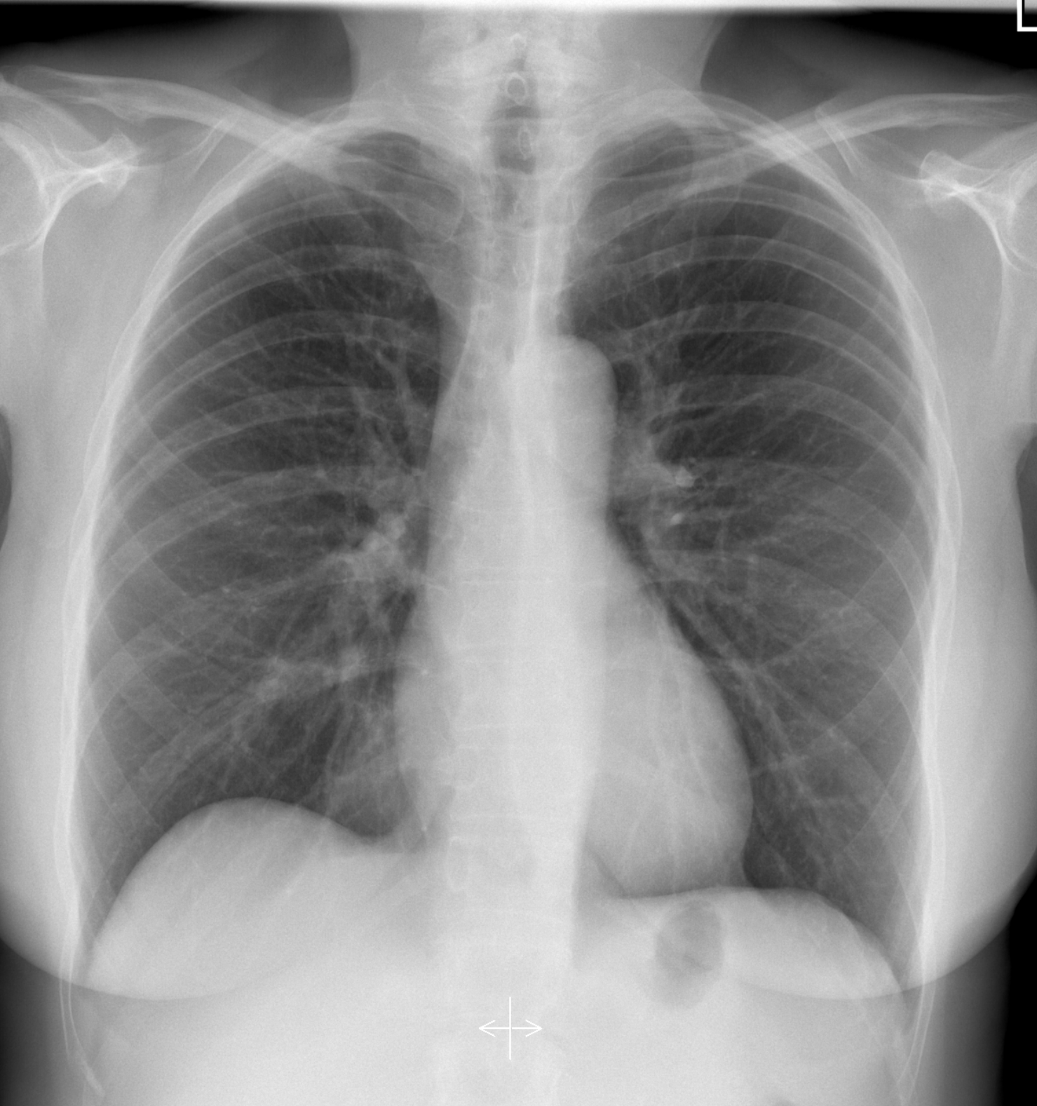
Understanding Thoracic X-Ray: A Comprehensive Guide
Thoracic X-ray is a vital diagnostic tool used in medical practice to evaluate the chest's anatomical structures and identify potential abnormalities. In this article, we will delve into the significance, procedures, interpretations, and common conditions detected through thoracic X-rays. Understanding how this imaging technique works can greatly enhance our approach to diagnosing and managing various respiratory and cardiovascular conditions.
The thoracic X-ray provides a non-invasive way to visualize the chest cavity, making it an essential procedure in both emergency and routine medical assessments. By capturing detailed images of the lungs, heart, and surrounding structures, healthcare professionals can make informed decisions regarding patient care. As we proceed, we will explore the critical aspects of thoracic X-ray, its benefits, limitations, and the role it plays in modern medicine.
This comprehensive guide aims to equip you with the knowledge required to understand thoracic X-rays better. Whether you are a medical professional, a student, or simply someone interested in healthcare, this article will shed light on the importance of this diagnostic tool and how it contributes to patient outcomes.
Table of Contents
- What is Thoracic X-Ray?
- Importance of Thoracic X-Ray
- How Thoracic X-Ray Works
- Preparation for Thoracic X-Ray
- Interpreting Thoracic X-Ray
- Common Conditions Detected by Thoracic X-Ray
- Limitations of Thoracic X-Ray
- Future of Thoracic Imaging
What is Thoracic X-Ray?
A thoracic X-ray is a diagnostic imaging technique that uses electromagnetic radiation to create images of the chest area, including the heart, lungs, blood vessels, and bones. This imaging method is widely used for assessing a variety of conditions, including pneumonia, heart failure, and lung tumors. The procedure is quick, painless, and requires minimal preparation.
Importance of Thoracic X-Ray
Thoracic X-rays hold immense significance in the medical field for several reasons:
- Early Detection: They can identify issues early, allowing for timely intervention.
- Non-invasive: The procedure is safe and requires no incisions.
- Cost-effective: Compared to other imaging techniques, thoracic X-rays are relatively inexpensive.
- Accessibility: Most medical facilities have the necessary equipment for performing X-rays.
How Thoracic X-Ray Works
Thoracic X-rays work by passing a controlled amount of radiation through the body. The radiation is absorbed differently by various tissues, creating a contrast that is recorded on a film or digital detector. The resulting image allows healthcare professionals to assess the condition of the chest structures.
Procedure Steps:
- The patient stands in front of the X-ray machine.
- A lead apron may be placed over the abdomen to protect from unnecessary radiation.
- The technician instructs the patient to take a deep breath and hold it while the image is captured.
- Multiple views may be taken to provide a comprehensive evaluation.
Preparation for Thoracic X-Ray
Preparation for a thoracic X-ray is straightforward:
- No special preparation is typically required.
- Patients should inform their healthcare provider if they are pregnant or suspect they might be.
- Jewelry or metal objects may need to be removed to prevent interference with the imaging.
Interpreting Thoracic X-Ray
Interpreting a thoracic X-ray requires specialized training. Radiologists examine the images for any abnormalities, such as:
- Enlarged heart
- Fluid in the lungs
- Pneumonia or other infections
- Bone fractures or abnormalities
Key Indicators:
- Look for symmetry in lung fields.
- Assess heart size and shape.
- Identify any abnormal shadows or densities.
Common Conditions Detected by Thoracic X-Ray
Thoracic X-rays can reveal various conditions, including:
- Pneumonia: Inflammation of the lungs.
- Congestive Heart Failure: Fluid buildup in the lungs.
- Lung Cancer: Abnormal masses in the lung tissue.
- Pneumothorax: Air in the pleural cavity.
Limitations of Thoracic X-Ray
While thoracic X-rays are invaluable, they do have limitations:
- They may not detect small tumors or early-stage diseases.
- Overlapping structures can obscure important details.
- Radiation exposure, though minimal, is a consideration, especially in young patients.
Future of Thoracic Imaging
The future of thoracic imaging is promising, with advancements in technology leading to improved diagnostic capabilities:
- Digital imaging techniques enhance image clarity.
- Artificial intelligence is being integrated to assist in image analysis.
- Hybrid imaging methods, such as PET-CT scans, provide more comprehensive evaluations.
Conclusion
In conclusion, thoracic X-rays are essential diagnostic tools that facilitate the early detection and management of various chest-related conditions. By understanding the significance, procedure, and interpretation of thoracic X-rays, both healthcare professionals and patients can engage in more informed discussions about health and treatment options. We encourage readers to consult with medical professionals for personalized advice and to share this article with others who may find the information valuable.
Call to Action
If you found this article helpful, please consider leaving a comment below or sharing it with your friends and family. For more informative articles on healthcare and medical imaging, explore our website further!
Final Thoughts
Thank you for taking the time to read this comprehensive guide on thoracic X-ray. We hope it enhances your understanding and piques your interest in the fascinating world of medical imaging. We look forward to welcoming you back for more insightful content in the future!
Article Recommendations
- Yvonne Elliman
- Bobby Charlton
- Unabated
- Holiday Inn Express Pcb
- Anant Real Name
- Nintendo Store Nyc
- How To Turn Off Scientific Notation In Excel
- Matt Dillon Wife
- Barron Trump Height In Feet
- Squealers Restaurant


